
Research
Neuroanatomy of the Drosophila Larva
Motivation
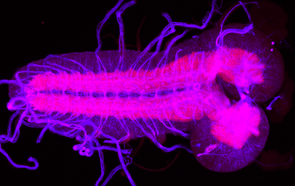
With only about 10,000 neurons, the central nervous system of the Drosophila larva is small enough to allow an anatomical reconstruction of all neurons and their connectivity. At the same time the larva and its behaviour are already complex enough to be of interest for neuroethology, and Drosophila is arguably the most important model organism in biology.
Scientific Questions
For neurobiological research on the larval Drosophila brain we develop methods for the evaluation of light and electron microscopy data as well as of the 3D neuron structures reconstructed from the latter (“neuron skeletons”).
In particular, we have created a standard brain (central nervous system) of the Drosophila larva from light microscopy images, and our software larvalign enables the registration of new scans into the coordinate system of the standard brain (Muenzing et al., 2018). We are also working on a multimodal registration between light and electron microscopy brain images.
At the level of the reconstructed individual neurons we have developed analysis and visualisation tools for neuroanatomical data evaluation: topologically correct but simplified representations of the 3D neuron structure in the form of 2D dendrograms allow a clear depiction of morphologically complex neurons and their connectivity in local networks (Strauch et al., 2018, Schleyer et al., 2020).
Partners
- Prof. Dr. Andreas Stephan Thum, Institute of Biology, University of Leipzig
- Dr. Katja Bühler, VRVis Research Center for Virtual Reality and Visualization, Vienna
- Prof. Dr. Volker Hartenstein, Department of Molecular, Cell and Developmental Biology, University of California, Los Angeles
External Funding
- DFG Research Grant, “The larval 4D standard brain of Drosophila melanogaster analysed at single-cell level”, Projektnr. 266382180
- DFG Research Grant, “Larvalbrain2.0: a research platform for the larval brain of Drosophila melanogaster”, project no. 441181781