Home/Research/Biomedical Image and Data Analysis/Synthesis, Segmentation, and Tracking of Live Cell Microscopy Image Data

Research

Synthesis, Segmentation, and Tracking of Live Cell Microscopy Image Data
Motivation

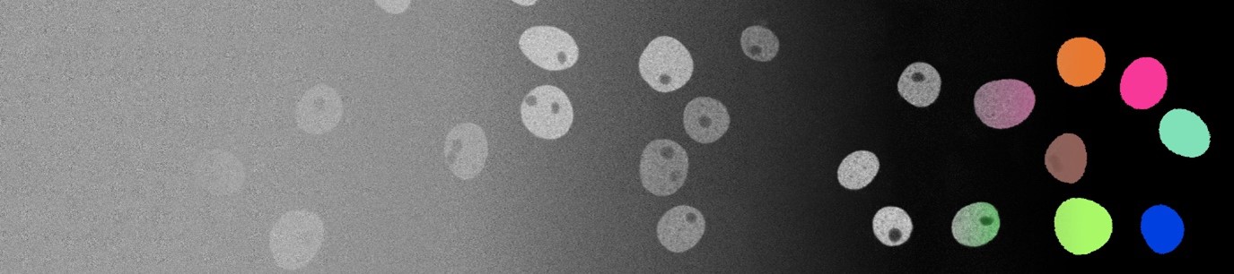
Developmental biology focuses on studying the processes of how cells migrate, interact and divide. Specifically, cell location and morphology, analyzed by segmentation and tracking approaches, constitute one of the key tasks in understanding the underlying principles of development. Technological advances allow biologists to capture and store extensive amounts of highly detailed microscopy image data, which renders the application of automated approaches essential. However, the potential of recent deep learning-based approaches is severely limited by the scarcity of fully annotated image data sets. Therefore, this project focuses on synthesis of fully annotated microscopy video data sets, eliminating the need for time-consuming manual annotation efforts.


Scientific Questions

  • How to generate realistic fully synthetic cell textures and shapes based on a limited amount of annotated real data?
  • How to capture the spatiotemporal relations of cell bodies and reflect this on the generated data?
  • How to make sure that there is enough variability in the synthetic data set and assess its quality?
  • How much improvement can we get by training segmentation and tracking models on abundant synthetic data as opposed to training them with limited real data?


Partners

External Funding

  • DFG Research Grant, "On-the-fly Data Synthesis for a Deep Learning-Based Analysis of 3D+t Microscopy Experiments", Project No. 447699143

Contact
Publications

  • 2024article

    Dennis Eschweiler, Rüveyda Yilmaz, Matisse Baumann, Ina Laube, Rijo Roy, Abin Jose, Daniel Brückner and Johannes Stegmaier

    Denoising Diffusion Probabilistic Models for Generation of Realistic Fully-Annotated Microscopy Image Data Sets

    In: PLOS Computational Biology 20 (2), S. e1011890

  • 2022inbook

    D. Eschweiler, I. Laube and J. Stegmaier

    Spatiotemporal Image Generation for Embryomics Applications

    In: Biomedical Image Synthesis and Simulation: Methods and Applications, S. (in press)

  • 2022inproceedings

    Dennis Eschweiler, Justus Schock and Johannes Stegmaier

    Probabilistic Image Diversification to Improve Segmentation in 3D Microscopy Image Data

    In: MICCAI International Workshop on Simulation and Synthesis in Medical Imaging (SASHIMI)

  • 2021article

    Dennis Eschweiler, Malte Rethwisch, Mareike Jarchow, Simon Koppers and Johannes Stegmaier

    3D fluorescence microscopy data synthesis for segmentation and benchmarking

    In: PLOS ONE 16 (12), S. 1-21, Public Library of Science

  • 2022inproceedings

    Dennis Eschweiler, Richard S. Smith and Johannes Stegmaier

    Robust 3D Cell Segmentation: Extending the View of Cellpose

    In: IEEE International Conference in Image Processing (ICIP)

  • 2021inproceedings

    C. Yang, D. Eschweiler and J. Stegmaier

    Semi-and Self-Supervised Multi-View Fusion of 3D Microscopy Images using Generative Adversarial Networks

    In: MICCAI Workshop on Machine Learning for Medical Image Reconstruction (MLMIR), S. (in press)

  • 2021inproceedings

    Dennis Eschweiler, Malte Rethwisch, Simon Koppers and Johannes Stegmaier

    Spherical Harmonics for Shape-Constrained 3D Cell Segmentation

    In: IEEE International Symposium on Biomedical Imaging (ISBI)

  • 2021inproceedings

    D. Eschweiler, A. Bhattacharyya, D. Moreno-Andrés, W. Antonin D. Bähr and J. Stegmaier

    CellCycleGAN: Spatiotemporal Microscopy Image Synthesis of Cell Populations using Statistical Shape Models and Conditional GANs

    In: IEEE International Symposium on Biomedical Imaging (ISBI)

  • 2019inproceedings

    Dennis Eschweiler, Tim Klose, Florian Nicolas Müller-Fouarge, Marcin Kopaczka and Johannes Stegmaier

    Towards Annotation-Free Segmentation of Fluorescently Labeled Cell Membranes in Confocal Microscopy Images

    In: MICCAI International Workshop on Simulation and Synthesis in Medical Imaging (SASHIMI), S. 81--89, Springer

  • 2019inproceedings

    Thiago V. Spina, Rohan C. Choudhury, Elliot Meyerowitz, Alexandre Cunha, Johannes Stegmaier Dennis Eschweiler

    CNN-based Preprocessing to Optimize Watershed-based Cell Segmentation in 3D Confocal Microscopy Images

    In: IEEE International Symposium on Biomedical Imaging (ISBI)

By continuing to use the site, you agree to the use of cookies. more information

English translation below.

Only your chosen language settings are stored in the cookies. Please also read our Privacy Policy.


The cookie settings on this website are set to "allow cookies" to give you the best browsing experience possible. If you continue to use this website without changing your cookie settings or you click "Accept" below then you are consenting to this.

Only your chosen language settings are stored in the cookies. Please also read our privacy policy.

Close