
Research
Image-based Phenotyping of Nematodes

Motivation
Nematodes (roundworms) form an extremely diverse animal phylum. Up to 4100 species of plant-parasitic nematodes are currently known. Many nematode species parasitise crops such as potatoes, sugar beet and soya beans and cause economic losses of up to 180 billion USD per year in agriculture worldwide. High-throughput analysis of nematodes from soil samples allows rapid quantification of nematode infestation as well as characterisation of the nematodes, thus advancing both cyst monitoring and resistance screening of plants.
At present, nematode cysts are picked out of organic residues by hand when soil samples are analysed, the amount of residue depending on the particular sample extraction method. Manual counting is time-consuming, and counting accuracy is affected by subjective decisions and by the human counter's ability to stay focused. At the same time, the main quantitative measurement of soil samples is limited to counting cysts, eggs and juveniles. Our aim is to extract more comprehensive phenotypic features in order to characterise the nematode population, which is likewise not possible with manual processing.
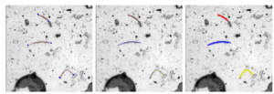
Abbildung 1: detection of endpoints, skeleton and segmentation of nematode juveniles
Scientific Questions
The overarching aim of this research project is to build a high-throughput system for the analysis of nematodes in soil samples, including nematode cysts, eggs and juveniles. A robust instance segmentation pipeline is the most important step, and a challenging one because of the heavily cluttered collection in the image. Besides optimising the segmentation pipeline, counting accuracy and the robustness of the extracted phenotypic features are also explicitly investigated.
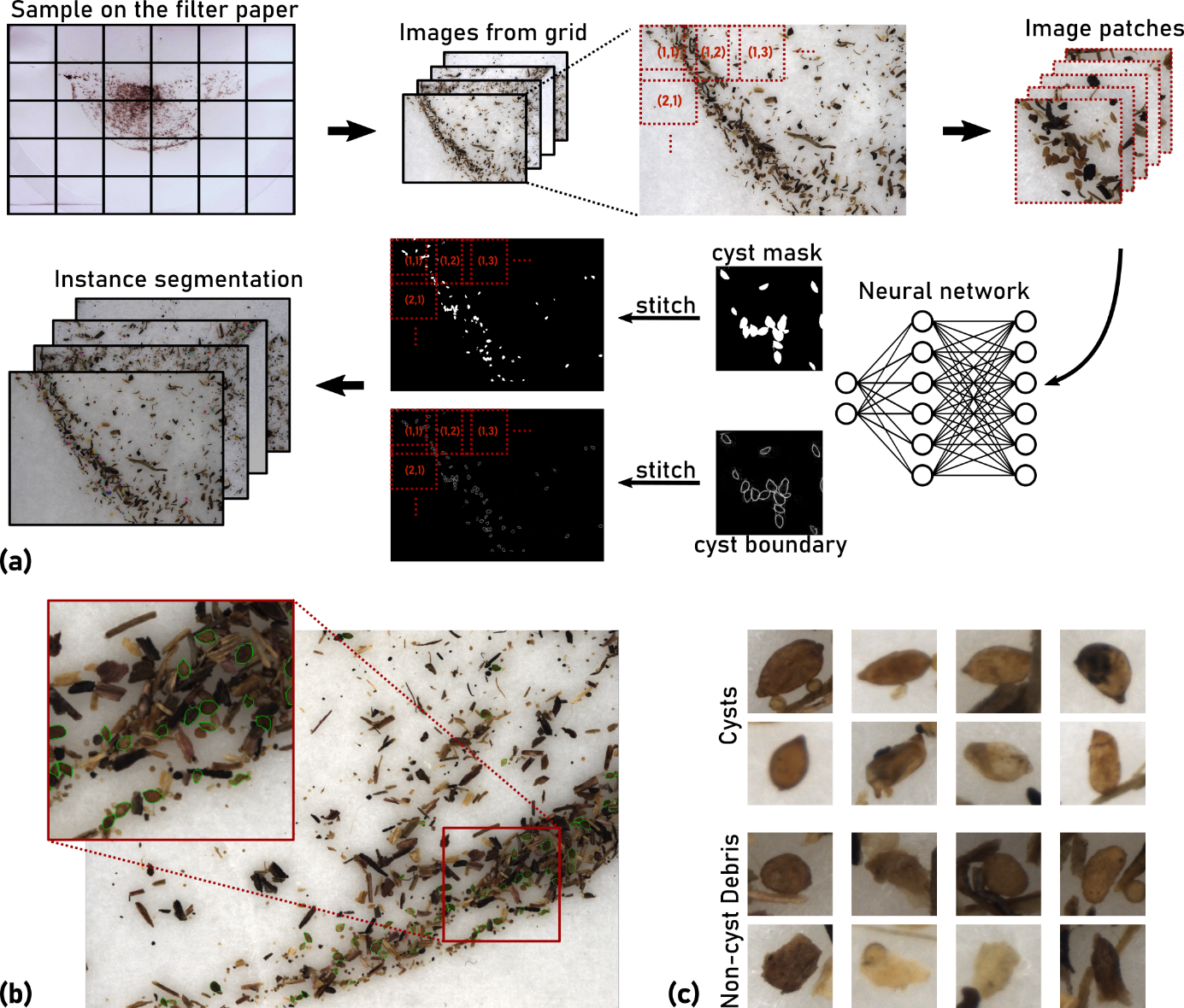
Abbildung 2: Image processing pipeline for the segmentation of nematode cysts. a) A high-resolution image is divided into tiles that are processed separately. A deep learning model predicts the cyst mask and the boundary. The boundary prediction is responsible for separating touching cysts. Detection of endpoints, skeleton and segmentation of nematode juveniles. b) Microscope image of a soil sample extract containing nematode cysts (marked in green) and organic debris particles. c) Examples of nematode cysts and debris particles resembling cysts.
Thesis Topics
New theses in the area of image-based phenotyping of nematodes are advertised regularly. In addition to the Overview , there are also numerous topics that have not yet been advertised, which will be gladly presented in a personal conversation.
Partners
- Dr. Matthias Daub, Julius Kühn Institute, Elsdorf
- Dr. Marcus Jansen, LemnaTec GmbH, Aachen
External Funding
- BMBF KMU-innovativ-19 project: “Phenotyping of nematodes with sensors: development of an automatic, image-based measurement method for nematodes at different developmental stages (PheNeSens)”, project no. FKZ: 031B0474C