Automated Analysis of Radiological Image Data
Motivation
Many diseases are diagnosed using medical imaging techniques of the domain of radiology. These include Computed Tomography (CT), Magnetic Resonance Imaging (MRI) as well as conventional radiographs (X-Ray). The diagnosis of the resulting images can only be done by clinical experts (radiologists) and is also very time intensive.
A (partially) automated evaluation could accelerate the diagnosis and greatly reduce the time between image acquisition, diagnosis and the start of therapy. Automated results could also be used as a “second opinion”. Particularly in emergencies or in the case of potentially life-threatening diseases, this offers the potential to greatly improve the therapy outcome.
Scientific Questions
In the field of medical processing and analysis of radiological image data there are four main tasks:
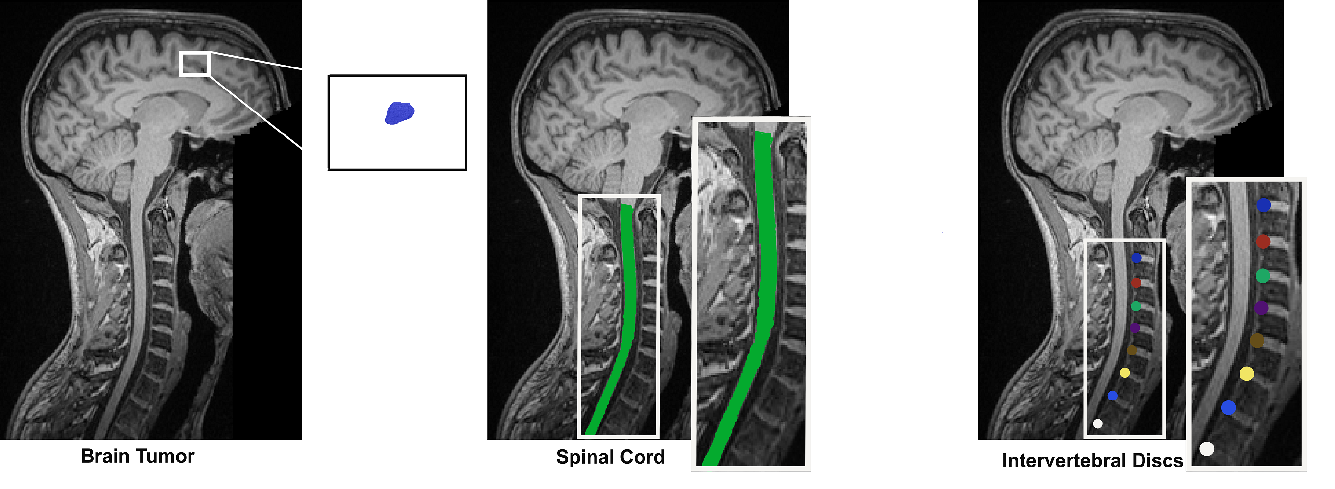
- Image classification places the image into one of several (categorical) classes. For example, in a given x-ray, whether or not there is a fractured leg.
- Image segmentation aims to provide masks for specific regions. In case of the x-ray it would be relevant to obtain masks for the different bones within a leg.
- Region of Interest Detection aims to provide a bounding-box surrounding the relevant portion of the image. Given the x-ray of a leg, this would be the exact location of the fracture.
- Measurements: For surgery planning and intervention planning in general, many different measures (usually distances and angles between well-defined anatomical points) are obtained manually by physicians. This is a very time-consuming work and has the potential of a great acceleration if replacing the manual effort of radiologists, orthopedics and surgeons by automated solutions.
All of these tasks suffer from the very limited amount of data that is available for medical domains in general and especially the rare amount of annotated data. Also, the three-dimensional nature of many of these imaging techniques make their processing much more challenging then corresponding two-dimensional data.
Theses
New theses are regularly advertised in the area of Automated Analysis of Radiological Image Data. In addition to the general overview, there are also numerous topics that have not yet been advertised, which will be gladly presented in a personal conversation.
Partners
- Prof. Dr. Fabian Kießling, Institute for Experimental Molecular Imaging (ExMI), RWTH Aachen
- Prof. Dr. Volmar Schulz, Institute for Experimental Molecular Imaging (ExMI), RWTH Aachen
- Dr. med. Sven Nebelung, Department for Diagnostic and Interventional Radiology, University Hospital RWTH Aachen
- Prof. Dr. med. Christinane Kuhl, Department for Diagnostic and Interventional Radiology, University Hospital RWTH Aachen
- Dr. med. Burkhard Gess, Department für Neurology, University Hospital RWTH Aachen
- Prof. Dr. med. Philippe Lambin, University of Maastricht, NL
External Funding
- DFG Research Grant, “More from less: Overcoming Data Scarcity for Deep Learning in Medical Image Computing”, Project nr. 455548460
- EURADIOMICS: The Euregional network for Distributed Deep Learning from Radiology imaging to improve decision making in Oncology
Contact
Publications
2022

Justus Schock, Yu-Chia Lan, Daniel Truhn, Marcin Kopaczka, Stefan Conrad, Sven Nebelung and Dorit Merhof
Monoplanar CT Reconstruction with GANs
In: IEEE International Conference on Image Processing Theory and Tools and Applications (IPTA)
2022
Reza Azad, Nika Khosravi and Dorit Merhof
SMU-Net: Style matching U-Net for brain tumor segmentation with missing modalities
In: 5th International Conference on Medical Imaging with Deep Learning (MIDL)
2021
Justus Schock, Daniel Truhn, Darius Nürnberger, Stefan Conrad, Marc S. Huppertz, Sebastian Keil, Christiane Kuhl, Dorit Merhof and Sven Nebelung
Artificial intelligence-based automatic assessment of lower limb torsion on MRI
In: Scientific Reports 11 (1)
2021
Reza Azad, Afshin Bozorgpour, Maryam Asadi-Aghbolaghi, Dorit Merhof and Sergio Escalera
Deep Frequency Re-Calibration U-Net for Medical Image Segmentation
In: ICCV Workshop on Computer Vision for Automated Medical Diagnosis (CVAMD)
2021
Michael Gadermayr, Lotte Heckmann, Kexin Li, Friederike Bähr, Madlaine Müller, Daniel Truhn, Dorit Merhof and Burkhard Gess
Image-to-Image Translation for Simplified MRI Muscle Segmentation
In: Frontiers in Radiology
2021
Michael Gadermayr, Maximilian Tschuchnig, Laxmi Gupta, Nils Krämer, Daniel Truhn, Dorit Merhof and Burkhard Gess
An asymmetric cycle-consistency loss for dealing with many-to-one mappings in image translation: a study on thigh MR scans
In: IEEE International Symposium on Biomedical Imaging (ISBI)
2020
Justus Schock, Marcin Kopaczka, Benjamin Agthe, Jie Huang, Paul Kruse, Daniel Truhn, Stefan Conrad, Gerald Antoch, Christiane Kuhl, Sven Nebelung and Dorit Merhof
A Method for Semantic Knee Bone and Cartilage Segmentation with Deep 3D Shape Fitting Using Data From the Osteoarthritis Initiative
In: MICCAI Workshop on Shape Analysis in Medical Imaging (ShapeMI)
2020
Christoph Haarburger, Gustav Müller-Franzes, Leon Weninger, Christiane Kuhl, Daniel Truhn and Dorit Merhof
Radiomics feature reproducibility under inter‐rater variability in segmentations of ct images
In: Scientific Reports (accepted)
2020
Christoph Haarburger, Justus Schock, Daniel Truhn, Philippe Weitz, Gustav Mueller-Franzes, Leon Weninger and Dorit Merhof
Radiomic Feature Stability Analysis based on Probabilistic Segmentations
In: IEEE International Symposium on Biomedical Imaging (ISBI)
2019
Michael Gadermayr, Kexin Li, Madlaine Müller, Daniel Truhn, Nils Krämer, Dorit Merhof and Burkhard Gess
Domain-specific data augmentation for segmenting MR images of fatty infiltrated human thighs with neural networks
In: Journal of Magnetic Resonance Imaging 49 (6)
2019
Christoph Haarburger, Nicolas Horst, Daniel Truhn, Mirjam Broeckmann, Simone Schrading, Christiane Kuhl and Dorit Merhof
Multiparametric Magnetic Resonance Image Synthesis using Generative Adversarial Networks
In: Eurographics Workshop on Visual Computing for Biology and Medicine (VCBM)
2019
Christoph Haarburger, Justus Schock, Michael Baumgartner, Oliver Rippel and Dorit Merhof
Delira: A High-Level Framework for Deep Learning in Medical Image Analysis
In: Journal of Open Source Software
2019
Christoph Haarburger, Michael Baumgartner, Daniel Truhn, Mirjam Broeckmann, Hannah Schneider, Simone Schrading, Christiane Kuhl and Dorit Merhof
Multi Scale Curriculum CNN for Context-Aware Breast MRI Malignancy Classification
In: International Conference on Medical Image Computing and Computer Assisted Intervention (MICCAI)
2019
Christoph Haarburger, Philippe Weitz, Oliver Rippel and Dorit Merhof
Image-based Survival Prediction for Lung Cancer Patients using CNNs
In: IEEE International Symposium on Biomedical Imaging (ISBI)
2018
Christoph Haarburger, Johannes Rüther, Daniel Truhn, Simone Schrading, Daniel Bug, Christiane K. Kuhl and Dorit Merhof
Abbreviated Breast Biopsy Procedure by Registration of Craniocaudal and Mediolateral Breast MR Images
In: IEEE International Symposium on Biomedical Imaging (ISBI)
2018
Christoph Haarburger, Peter Langenberg, Daniel Truhn, Hannah Schneider, Johannes Thüring, Simone Schrading, Christiane K. Kuhl and Dorit Merhof
Transfer Learning for Breast Cancer Malignancy Classification based on Dynamic Contrast-Enhanced MR Images
In: Bildverarbeitung für die Medizin (BVM)
2018
Michael Gadermayr, Constantin Disch, Madlaine Müller, Dorit Merhof and Burkhard Gess
A Comprehensive Study on Automated Muscle Segmentation for Assessing Fat Infiltration in Neuromuscular Diseases
In: Magnetic Reconance Imaging (MRI) 48
Automated Analysis of Radiological Image Data
Motivation
Many diseases are diagnosed using medical imaging techniques of the domain of radiology. These include Computed Tomography (CT), Magnetic Resonance Imaging (MRI) as well as conventional radiographs (X-Ray). The diagnosis of the resulting images can only be done by clinical experts (radiologists) and is also very time intensive.
A (partially) automated evaluation could accelerate the diagnosis and greatly reduce the time between image acquisition, diagnosis and the start of therapy. Automated results could also be used as a “second opinion”. Particularly in emergencies or in the case of potentially life-threatening diseases, this offers the potential to greatly improve the therapy outcome.
Scientific Questions
In the field of medical processing and analysis of radiological image data there are four main tasks:
- Image classification places the image into one of several (categorical) classes. For example, in a given x-ray, whether or not there is a fractured leg.
- Image segmentation aims to provide masks for specific regions. In case of the x-ray it would be relevant to obtain masks for the different bones within a leg.
- Region of Interest Detection aims to provide a bounding-box surrounding the relevant portion of the image. Given the x-ray of a leg, this would be the exact location of the fracture.
- Measurements: For surgery planning and intervention planning in general, many different measures (usually distances and angles between well-defined anatomical points) are obtained manually by physicians. This is a very time-consuming work and has the potential of a great acceleration if replacing the manual effort of radiologists, orthopedics and surgeons by automated solutions.
All of these tasks suffer from the very limited amount of data that is available for medical domains in general and especially the rare amount of annotated data. Also, the three-dimensional nature of many of these imaging techniques make their processing much more challenging then corresponding two-dimensional data.
Theses
New theses are regularly advertised in the area of Automated Analysis of Radiological Image Data. In addition to the general overview, there are also numerous topics that have not yet been advertised, which will be gladly presented in a personal conversation.
Partners
- Prof. Dr. Fabian Kießling, Institute for Experimental Molecular Imaging (ExMI), RWTH Aachen
- Prof. Dr. Volmar Schulz, Institute for Experimental Molecular Imaging (ExMI), RWTH Aachen
- Dr. med. Sven Nebelung, Department for Diagnostic and Interventional Radiology, University Hospital RWTH Aachen
- Prof. Dr. med. Christinane Kuhl, Department for Diagnostic and Interventional Radiology, University Hospital RWTH Aachen
- Dr. med. Burkhard Gess, Department für Neurology, University Hospital RWTH Aachen
- Prof. Dr. med. Philippe Lambin, University of Maastricht, NL
External Funding
- DFG Research Grant, “More from less: Overcoming Data Scarcity for Deep Learning in Medical Image Computing”, Project nr. 455548460
- EURADIOMICS: The Euregional network for Distributed Deep Learning from Radiology imaging to improve decision making in Oncology
Contact
Publications
2022
Justus Schock, Yu-Chia Lan, Daniel Truhn, Marcin Kopaczka, Stefan Conrad, Sven Nebelung and Dorit Merhof
Monoplanar CT Reconstruction with GANs
In: IEEE International Conference on Image Processing Theory and Tools and Applications (IPTA)
2022
Reza Azad, Nika Khosravi and Dorit Merhof
SMU-Net: Style matching U-Net for brain tumor segmentation with missing modalities
In: 5th International Conference on Medical Imaging with Deep Learning (MIDL)
2021
Justus Schock, Daniel Truhn, Darius Nürnberger, Stefan Conrad, Marc S. Huppertz, Sebastian Keil, Christiane Kuhl, Dorit Merhof and Sven Nebelung
Artificial intelligence-based automatic assessment of lower limb torsion on MRI
In: Scientific Reports 11 (1)
2021
Reza Azad, Afshin Bozorgpour, Maryam Asadi-Aghbolaghi, Dorit Merhof and Sergio Escalera
Deep Frequency Re-Calibration U-Net for Medical Image Segmentation
In: ICCV Workshop on Computer Vision for Automated Medical Diagnosis (CVAMD)
2021
Michael Gadermayr, Lotte Heckmann, Kexin Li, Friederike Bähr, Madlaine Müller, Daniel Truhn, Dorit Merhof and Burkhard Gess
Image-to-Image Translation for Simplified MRI Muscle Segmentation
In: Frontiers in Radiology
2021
Michael Gadermayr, Maximilian Tschuchnig, Laxmi Gupta, Nils Krämer, Daniel Truhn, Dorit Merhof and Burkhard Gess
An asymmetric cycle-consistency loss for dealing with many-to-one mappings in image translation: a study on thigh MR scans
In: IEEE International Symposium on Biomedical Imaging (ISBI)
2020
Justus Schock, Marcin Kopaczka, Benjamin Agthe, Jie Huang, Paul Kruse, Daniel Truhn, Stefan Conrad, Gerald Antoch, Christiane Kuhl, Sven Nebelung and Dorit Merhof
A Method for Semantic Knee Bone and Cartilage Segmentation with Deep 3D Shape Fitting Using Data From the Osteoarthritis Initiative
In: MICCAI Workshop on Shape Analysis in Medical Imaging (ShapeMI)
2020
Christoph Haarburger, Gustav Müller-Franzes, Leon Weninger, Christiane Kuhl, Daniel Truhn and Dorit Merhof
Radiomics feature reproducibility under inter‐rater variability in segmentations of ct images
In: Scientific Reports (accepted)
2020
Christoph Haarburger, Justus Schock, Daniel Truhn, Philippe Weitz, Gustav Mueller-Franzes, Leon Weninger and Dorit Merhof
Radiomic Feature Stability Analysis based on Probabilistic Segmentations
In: IEEE International Symposium on Biomedical Imaging (ISBI)
2019
Michael Gadermayr, Kexin Li, Madlaine Müller, Daniel Truhn, Nils Krämer, Dorit Merhof and Burkhard Gess
Domain-specific data augmentation for segmenting MR images of fatty infiltrated human thighs with neural networks
In: Journal of Magnetic Resonance Imaging 49 (6)
2019
Christoph Haarburger, Nicolas Horst, Daniel Truhn, Mirjam Broeckmann, Simone Schrading, Christiane Kuhl and Dorit Merhof
Multiparametric Magnetic Resonance Image Synthesis using Generative Adversarial Networks
In: Eurographics Workshop on Visual Computing for Biology and Medicine (VCBM)
2019
Christoph Haarburger, Justus Schock, Michael Baumgartner, Oliver Rippel and Dorit Merhof
Delira: A High-Level Framework for Deep Learning in Medical Image Analysis
In: Journal of Open Source Software
2019
Christoph Haarburger, Michael Baumgartner, Daniel Truhn, Mirjam Broeckmann, Hannah Schneider, Simone Schrading, Christiane Kuhl and Dorit Merhof
Multi Scale Curriculum CNN for Context-Aware Breast MRI Malignancy Classification
In: International Conference on Medical Image Computing and Computer Assisted Intervention (MICCAI)
2019
Christoph Haarburger, Philippe Weitz, Oliver Rippel and Dorit Merhof
Image-based Survival Prediction for Lung Cancer Patients using CNNs
In: IEEE International Symposium on Biomedical Imaging (ISBI)
2018
Christoph Haarburger, Johannes Rüther, Daniel Truhn, Simone Schrading, Daniel Bug, Christiane K. Kuhl and Dorit Merhof
Abbreviated Breast Biopsy Procedure by Registration of Craniocaudal and Mediolateral Breast MR Images
In: IEEE International Symposium on Biomedical Imaging (ISBI)
2018
Christoph Haarburger, Peter Langenberg, Daniel Truhn, Hannah Schneider, Johannes Thüring, Simone Schrading, Christiane K. Kuhl and Dorit Merhof
Transfer Learning for Breast Cancer Malignancy Classification based on Dynamic Contrast-Enhanced MR Images
In: Bildverarbeitung für die Medizin (BVM)
2018
Michael Gadermayr, Constantin Disch, Madlaine Müller, Dorit Merhof and Burkhard Gess
A Comprehensive Study on Automated Muscle Segmentation for Assessing Fat Infiltration in Neuromuscular Diseases
In: Magnetic Reconance Imaging (MRI) 48


