PET/MRI System Development
Motivation
Positron emission tomography (PET) is a highly sensitive medical imaging modality with broad application, e.g. in oncology for tumor diagnosis. PET is based on functional tracers injected into the patient and accumulating at their specific target structures in the body after a certain time. During the radioactive decay of the tracers, they emit positrons that annihilate with electrons. This produces two oppositely oriented, high-energy gamma photons that leave the body and are registered by a surrounding ring of detectors
The detectors consist of passive and active components. Scintillation crystals stop the gamma photons and convert them into several thousand, mostly bluish, optical photons. These are recorded, digitized, and processed by optical sensors. The system software then uses calibration and special algorithms to calculate the time of the gamma interaction, the deposited energy, and the location of the interaction from the raw data
Two corresponding, detected events result in a so-called line-of-response (LOR). Around 100 million such LORs are used to reconstruct the tracer distribution in the body, making the functional processes visible. By precisely determining the time difference in the arrival of the two gamma-ray photons (time-of-flight), the place of origin along the LOR can be localized even more precisely. This additional information significantly contributes to improving image quality.
For diagnostic purposes, PET is often combined with anatomical imaging such as computed tomography (CT) or magnetic resonance imaging (MRI). MRI offers the advantage of a higher soft tissue contrast and a radiation-free examination. For this reason, medical technology companies such as Siemens, GE, and Philips have developed PET/MRI systems that image the entire body and are particularly suitable for addressing oncological issues.
Our research group has many years of experience in building MRI-compatible PET research systems. These systems allow us to answer specific questions much more precisely than is possible with commercial devices. We work closely with a wide range of industrial and academic partners as well as users to develop innovative solutions for current scientific and clinical challenges.
The spin-off Hyperion Hybrid Imaging Systems GmbH [1] is a partner in several research projects and is also driving the use of the MRI-compatible PET platform.


Research Topics
Our research and development activities cover the entire imaging chain: from the tracer decay to the detection of gamma photons, analog and digital signal processing, data acquisition and processing with high-performance computing concepts, and image reconstruction. In doing so, we work on a wide range of physics issues, including the simulation, development, and characterization of systems and detectors. Examples of finalized systems are the HYPMED PET/MRT breast scanner (s. Figure 1), the split-gradient PET/MRT scanner (s. Figure 2), and the neuro PET/MRI Insert (s. Figure 3).
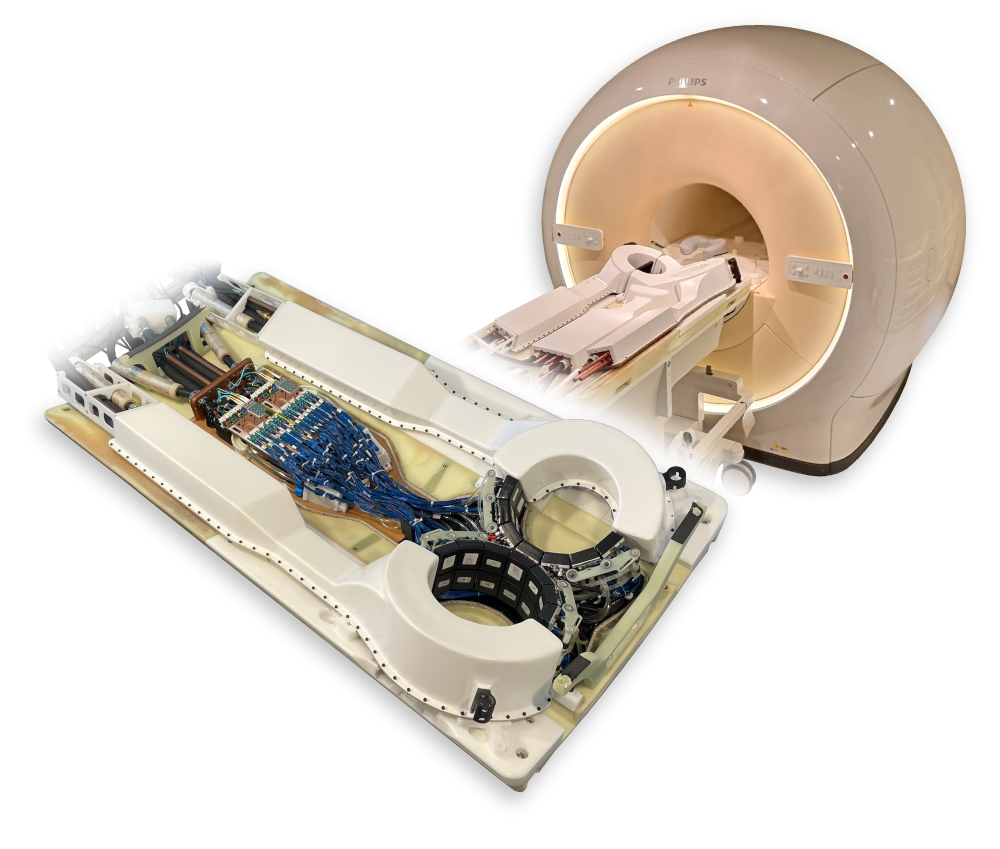
Figure 1: The EU project HYPMED [2] developed a PET insert for a 1.5 T MRI that enables simultaneous imaging of both breasts with MRI and PET. The HYPMED hybrid scanner is designed to provide high-resolution images for improved breast cancer diagnosis and early detection.
In addition, there is a focus on the development and optimization of electronics and firmware as well as on the design of innovative software solutions. We utilize state-of-the-art machine learning and high-performance processing methods to further improve image quality and imaging efficiency. For example, Gradient Tree Boosting (GTB) is used in the HYPMED scanner to accurately predict a 3D gamma interaction position from the read-out 2D light distributions of the optical photons. The influence of the algorithms can be evaluated individually per detector or on a PET image basis. Our platform also allows the implementation of various algorithms from libraries such as PyTorch and xgboost.
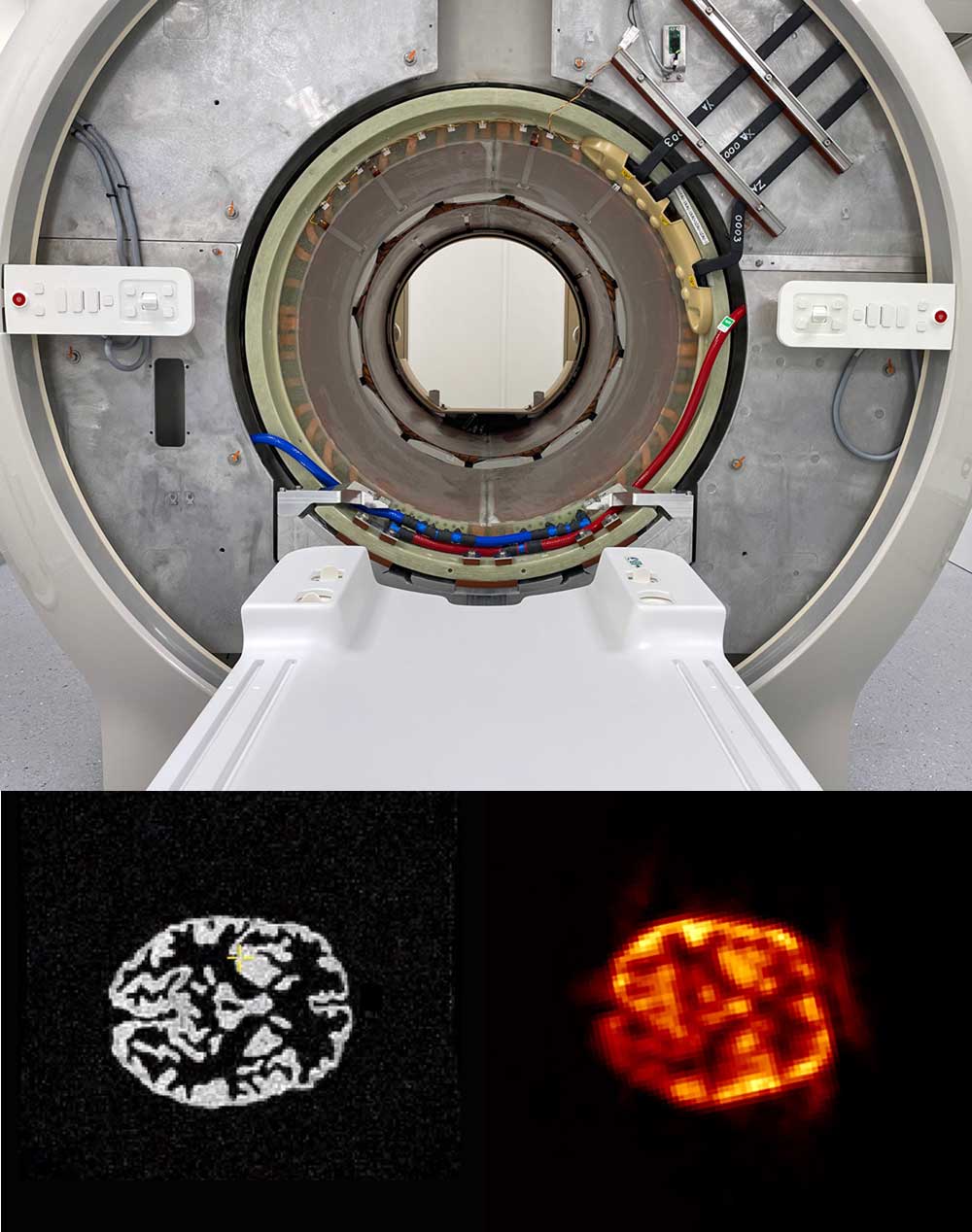
Figure 2: In cooperation with Hyperion Hybrid Imaging Systems GmbH, Philips HealthCare, Futura Composites and UMC Utrecht, a 1.5 T MRI was modified in such a way that PET detectors were integrated in the gap of a split gradient. This means that the full inner diameter of the MRI is available for simultaneous imaging.
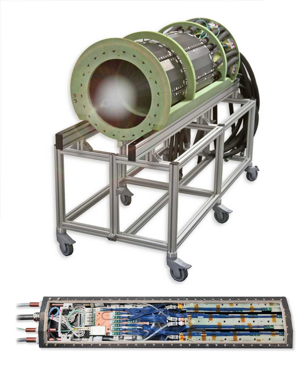
Figure 3: In cooperation with Forschungszentrum Jülich, Siemens Healthineers, Monash University, Inviscan and Affinity Imaging, a neuro-insert for a 7 T MRI was set up [4]. With an axial field of view of 25 cm and an image resolution of about 1.5 mm, the system is one of the most powerful in the world.
For our work, we have exclusive access to a 1.5 Tesla clinical MRI system. We use these for MRI research as well as for detailed investigations of the interferences between PET components and MRI image acquisition. HD-MetaPET project funded by the German Federal Ministry of Education and Research (BMBF), we are developing a PET insert that enables simultaneous PET/MRI images. The aim is to combine the advantages of both modalities to open up new diagnostic possibilities. Part of the system development is simulation work with frameworks such as Geant4 and GATE, e.g., to optimize the placement of the individual PET detectors in the system for the highest possible performance under the boundary condition of MRI interference. Furthermore, new calibration methods are developed for the use of machine learning techniques at the system level. These include gamma interaction positioning (Y. Kuhl, 2024), as well as time calibration (S. Naunheim, 2023).
Another BMBF-funded project, S-PET, is dedicated to the development of a novel PET detector. This detector is specifically designed to meet the requirements of neurological imaging and is intended to support research and diagnosis in the field of Alzheimer’s disease. A central component is the development of compression schemes that reduce the number of optical sensors, optimized for the detector performance parameters of time, energy, and positioning resolution.
Vacancies
Within the framework of the HD-MetaPET and S-PET projects, PhD positions to fill.
Thesis Topics
We offer various theses along the entire PET image creation chain. These can be tailored to the candidate profile in the direction of electronics and firmware development, laboratory, and characterization work or with a focus on software development, data analysis, and algorithm development.
Current topics can be found here. .
Compression of PET-Data Contact Tim Claßen
More information about the research project
Various Topics in Positron-Emission-Tomography: From Scanner Calibration To Image Reconstruction Contact Julian Thull
More information about the research project
References
[1] https://hyperion-his.com/
[2] https://www.cbmb.ukaachen.de/hypmed-brustkrebs-forschungsprojekt/
[3] https://www.umcutrecht.nl/nieuws/deel-mri-pet-geinstalleerd-the-next-step?lang=en
[4] https://www.fz-juelich.de/de/inm/inm-4/forschungsgruppen/pet/technische-aspekte-der-mr-pet/brainpet-7t
[5] https://www.exmi.rwth-aachen.de/cms/exmi/forschung/forschungsprojekte/~baxolf/hd-metapet/