Immunocytochemical Pre-Screening of Stained Cell Smears
Background
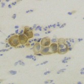
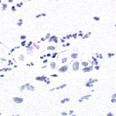
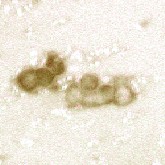
In recent years proteins within cells have been identified that are directly related to particular diseases, e.g. cancer. Many of these proteins can be visualized with immunocytochemical marker stains. For example cells infected by a virus can be visualized by attaching a stain to antibodies which in turn bind to a specific upregulated protein after virus infection. The antibody, docked to the affected cell, is stained by a chromogene which then can be identified in a microscopic inspection. Human papilloma viruses (HPV), e.g., are responsible for almost all cervical cancers, and the infection can be detected with the specific p16-antibody in cervical brush smears with high sensitivity.

Mission
An immunocytological investigation can therefore non-invasively identify high risk patients for this type of cancer. The automated scanning of slides followed by computer aided detection of marker-positive cells will help physicians to cope with the high load of specimens in a screening scenario and to reach a more reliable diagnostic result.


Publication
Research topics
Ahmet Aydin
Improving Specificity of Immunocytochemical Marker Detection via 40x Magnified Microscopic Images.
Tobias Wächter
Detection of stained vaginal mucus to reduce artifacts in immunocytochemical pre-screening.
(Detektion von eingefärbtem Vaginalschleim für die Artefakt-Reduktion im immunzytochemischen Pre-Screening)
Joschka zur Jacobsmühlen
Development of detection algorithms for immunocytochemical markers in microscopy images.
(Entwicklung von Detektionsverfahren für immunzytochemische Marker in Mikroskopie-Bildern)
Contact person:
- David Friedrich, Tel.: +49 (241) 80-27803
- Kraisorn Chaisaowong, Tel.: +49 (241) 80-27865
Medical Partner:
- Univ.-Prof. Dr. med. Ruth Knüchel-Clarke, Director
Dr. med. Till Braunschweig, Assistant physician - Institute of Pathology, (Institut für Pathologie, IfP),
University Hospital Aachen
– Medical Faculty RWTH –
Coordinator Partner:
- Univ.-Prof. i.R. Dr.-Ing. Rolf H. Jansen
Chair of Electromagnetic Theory of RWTH Aachen
Funded by:
- Exploratory Research Space @ RWTH Aachen (ERS)
- Pathfinder Project (Seed Fund): MedTec
- The project was supported by the excellence initiative of the German federal and state governments.

