Pleuramesothelioma
Computer-Assisted Diagnosis for Early Stage Pleural Mesothelioma: Automated Detection and Quantitative Assessment of Pleural Thickenings from Thoracic CT Images
Background
There was almost a century of import and consumption of asbestos in the industrialized countries in Western Europe and Northern America, instead of knowing the fibrotic and carcinogenic potential of the inhalation of asbestos fibers. As a matter of fact, malignant pleural mesothelioma, which is normally a rare tumor, was found thousand times more often in persons exposed regularly to asbestos during their daily work than in the normal population. Today, it is statistically documented that 70-90% of malignant pleural mesotheliomata are traced back to asbestos exposure. At least three decades elapsed, until it came to a statutory prohibition, for instance in the year 1993 in Germany. There are several types of asbestos, differentiated by their filament forms. The narrow and needle-like type (amphiboles such as crocidolite and amosite, the so-called blue asbestos) was classified to be more carcinogenic than those that are curled and pliable (chrysotile, the so-called white asbestos), which is biologically more degradable and less harmful. Both types were widely applied in various industrial branches.
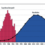
Because of the industrialization after the Second World War, use of asbestos reached its maximum during the late 1970s for example in Germany. Due to a long latency period of – on the average – 35 years (range between 10 to 60 years) after asbestos exposure, occurrence of malignant pleural mesothelioma morbidity and mortality in Germany is expected to peak during 2010s. Worldwide, asbestos related deaths are expected to rise to at least a million over the next 30 years.
At present in Germany, an assessment program is applied to the asbestos exposed group of persons. This assessment program includes thoracic CT imaging for non-invasive diagnostics in high risk groups. Physicians usually have to manually investigate over 80 layers of thoracic images to find all kinds of pleural or pericardial thickenings. After diagnosis of malignant pleural mesothelioma, survival time was documented to be between 4 to 18 months without any therapy. For early stage pleural mesothelioma however, pleurectomy together with perioperative treatment can reduce the morbidity and delay the mortality. Thus, efficient and reliable diagnosis of early stage pleural mesothelioma is a key factor to ease the consequences of the expected peak of malignant pleural mesothelioma.
 |
 |
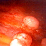
| Thoracoscopy of pleural mesotheliomata | The expected burden of pleural mesothelioma morbidity in Germany with peak of maximal 1000 cases in the year 2017 |
Vision
A typical mesothelioma related clinical non-invasive diagnosis is based on thoracic axial CT images. Depending on the layers’ thickness, the number of images varies between 80 slices with a thickness of 5 mm to about 700 slices with a thickness of 0.5 mm. Physicians view each slice on a workstation in order to find pleural thickenings. A simultaneous second diagnostic aim is the occurrence of lung nodules. The diagnostic findings are documented in a standardized form containing data such as their size, position, and growth rate. This visual diagnostic approach is a very time consuming procedure, taking about 20 to 30 minutes per data set, and this is considered as often being subjective. Indeed, studies have shown that the inherent complexity of tumor measurements impair an accurate assessment. Furthermore, differences in the diagnostic results were found between different investigating clinicians (inter-reader variability). To increase the accuracy of the localization and of the topological information of these quite small image regions within a subjective visual evaluation, an even longer investigating time would be needed for each data set. However, due to the increasing number of investigations and high work load of the physicians involved, this solution is not practicable. Therefore, a method is needed to provide a more accurate assessment of pleural mesothelioma at an early stage, which is reliable, consistent, and reproducible.
Mission
The aim of this work is to develop a computer system to automatically detect and quantitatively assess pleural thickenings in axial thoracic CT images. In case of a malignant finding it is important to consider that the earlier neoplasms are found, the higher the survival median of asbestos patients can be expected. For the therapy after a positive diagnosis of pleural mesothelioma, an accurate and reliable documentation of the evolution over time, such as growth rate, is essential.
Test session runs on real data set provided reliable and reproducible results.
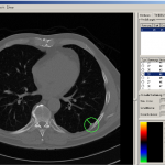
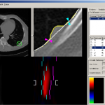
| Graphical User Interface of the System PleuraDatPlus | |
 |
 |
| Finding of a candidate pleural thickening. | Characteristics’ assessment. |
Our system is currently transferred into routine clinical use.
| Contact: | Peter Faltin, Tel.: +49 (241) 80-27865 |
| Partners: | Univ.-Prof. Dr. med. Thomas Kraus |
| Institut für Arbeitsmedizin und Sozialmedizin, | |
| Universitätsklinikum Aachen, AÖR, | |
| – Medizinische Fakultät der RWTH – | |
| Funding: | Deutsche Gesetzliche Unfallversicherung (DGUV) |
| Projekt-Nr. FF-FB0148 | |
| Last update: | 02.03.2011 |

