Classification of colon polyps for early cancer detection
Supported by Bundesministerium fuer Bildung und Forschung within the Innovationswettbewerb Medizintechnik 2009
Colorectal cancer
Colorectal cancer is one of the leading causes of death in the western countries. In 2004 about 20.000 patients died from malignant lesions of the colon in Germany, and approximately every fifth human develops colorectal cancer in the course of his life. The best chance for survival is achieved with an early detection of pre-cancer states since a successful therapy often depends on whether the cancer has started to developed metastases or not.
Early diagnosis
There are several kinds of screening methods. Starting at the age of 50 patients are advised to have their stool tested for traces of blood. Occult (hidden) blood may be an indication for colorectal cancer or a benign inflammatory bowel disease. A positive result necessitates a more thorough inspection, usually a colonoscopy, either to rule out colorectal cancer or to allow an early detection.
Due to the age-based risk of colorectal cancer, it is recommended for patients at the age of 55 or above to have regular colonoscopy screenings. During colonoscopy the colon is inspected by a medical practitioner using a flexible endoscope inserted into the rectum. Thus, it is possible to view the inner colon surface on a screen in the surgery. The medical practitioner can inspect the colon and detect alterations of the tissue.
Conservative treatment
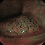
Next to established methods like the amputation and subsequent histological analysis of suspicious tissue there are new methods for the classification of colon polyps. One example is chromo endoscopy during which the surface structure of the mucosa is visualized by the application of chromatic dye. Another method is task-specific illumination (NBI, Narrow Band Imaging), which can point out surface structure and even underlying blood vessels. A classification based on the highlighting of the surface structure and potentially present blood vessels is feasible without a removal and histological analysis of the tissue. This method is especially conservative in case of a negative result as benign polyps do not necessarily have to be removed.
 |
 |
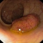
| Colorectal polyp under standard illumination. | Colorectal polyp under NBI illumination. Bleedings on the polyp surface are easily detectable as dark spots |
The detection rate of this method depends on the investigator and her/his knowledge of the field. The Institute of Imaging & Computer Vision, RWTH Aachen University, in cooperation with Internal Medicine III, RWTH Aachen University Hospital, currently researches methods to make objective and reproducible evaluations supporting the medical practitioner in making the final decision. Several methods of texture classification and blood vessel identification are under investigation.
Contact
Sebastian Groß, Tel.: +49 (241) 80-27862

