
Bildbasierte Phänotypisierung von Nematoden
Motivation
Nematoden (Fadenwürmer) bilden einen äußerst vielfältigen Tierstamm. Derzeit sind bis zu 4100 Arten von pflanzenparasitären Nematoden bekannt. Viele Nematodenarten parasitieren auf Nutzpflanzen wie Kartoffeln, Zuckerrüben und Sojabohnen und verursachen in der Landwirtschaft weltweit jährlich wirtschaftliche Verluste von bis zu 180 Mrd. USD. Eine Hochdurchsatzanalyse von Nematoden aus Bodenproben ermöglicht eine schnelle Quantifizierung des Nematodenbefalls sowie die Charakterisierung der Nematoden und bringt damit sowohl die Zystenüberwachung als auch das Resistenzscreening von Pflanzen voran.
Derzeit werden die Nematodenzysten bei der Analyse von Bodenproben von Hand aus organischen Rückständen herausgepickt, wobei die Menge der Rückstände von der jeweiligen Probenextraktionsmethode abhängt. Die manuelle Zählung ist zeitaufwändig, und die Zählgenauigkeit wird durch subjektive Entscheidungen und die Fähigkeit des menschlichen Zählers, konzentriert zu bleiben, beeinflusst. In der Zwischenzeit beschränkt sich die wichtigste quantitative Messung von Bodenproben auf die Zählung von Zysten, Eiern und Jungtieren. Unser Ziel ist es, umfassendere phänotypische Merkmale zu extrahieren, um die Nematodenpopulation zu charakterisieren, was durch manuelle Verarbeitung ebenfalls nicht möglich ist.
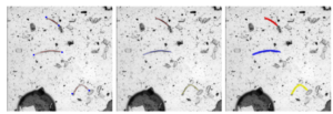
Abbildung 1: Erkennung von Endpunkten, Skelett und Segmentierung von Nematoden-Jungtieren
Wissenschaftliche Fragestellungen
Das übergeordnete Ziel dieses Forschungsprojekts ist der Aufbau eines Hochdurchsatzsystems für die Analyse von Nematoden in Bodenproben, einschließlich Nematodenzysten, Eiern und Jungtieren. Eine robuste Pipeline zur Segmentierung von Instanzen ist der wichtigste Schritt, der aufgrund der stark unübersichtlichen Sammlung im Bild eine Herausforderung darstellt. Neben der Optimierung der Segmentierungspipeline werden auch die Zählgenauigkeit und die Robustheit der extrahierten phänotypischen Merkmale explizit untersucht.
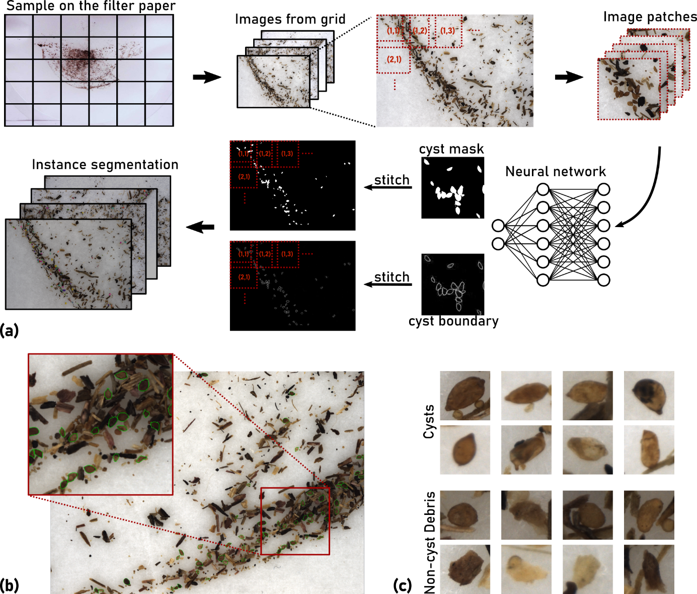
Abbildung 2: Bildverarbeitungspipeline für die Segmentierung von Nematodenzysten. a) Ein hochauflösendes Bild wird in Kacheln unterteilt, die separat verarbeitet werden. Ein Deep-Learning-Modell prognostiziert die Zystenmaske und die Begrenzung. Die Grenzvorhersage ist für die Trennung von sich berührenden Zysten verantwortlich. Erkennung von Endpunkten, Skelett und Segmentierung von Nematodenjungtieren. b) Mikroskopische Aufnahme eines Bodenprobenextrakts, der Nematodenzysten (grün markiert) und organische Trümmerpartikel enthält. c) Beispiele für Nematodenzysten und Trümmerpartikel, die den Zysten ähneln.
Abschlussarbeiten
Es werden regelmäßig neue Abschlussarbeiten im Bereich Bildbasierte Phänotypisierung von Nematoden ausgeschrieben. Neben der Gesamtübersicht gibt es auch noch zahlreiche, noch nicht ausgeschriebene Themen, die gerne im persönlichen Gespräch vorgestellt werden.
Kooperationspartner
- Dr. Matthias Daub, Julius Kühn Institut, Elsdorf
- Dr. Marcus Jansen, LemnaTec GmbH, Aachen
Drittmittel
- BMBF KMU-Innovativ-19 Projekt. “Phaenotypisierung von Nematoden mit Sensoren: Entwicklung einer automatischen, bildgestützten Messmethode für Neamtoden in verschiedenen Entwicklungsstadien (PheNeSens)” Projektnr. FKZ: 031B0474C
Kontakt

Veröffentlichungen

A CNN Framework Based on Line Annotations for Detecting Nematodes in Microscopic Images
In: IEEE International Symposium on Biomedical Imaging (ISBI)
