PET Image Reconstruction
Motivation
Positron-Emission-Tomography is a functional medical imaging technique with proven utility in fields as oncology, cardiology, and neurology. By using radioactive tracers, PET allows for the detailed visualization of physiological processes at the molecular level. Our research focuses on developing new reconstruction methods and improving image quality by improving scanner calibration and normalization.
Research Topics
PET scanners are highly complex systems in which image quality critically depends on both accurate system calibration and robust qunatitative image reconstruction. In practice, deviations from ideal system behaviour directly propagate into reconstruction artifacts and quantification errors. Our research treats calibration and reconstruction as a single, coupled problem with the goal of improving image quality under realistic imaging conditions.
In-System Calibration
Following assembly, PET systems typically deviate from their ideal geometric and physical configuration due to detector positioning errors, orientation mismatches, and variations in detector response. These effects influence gamma interaction positioning, timing, and energy estimation, ultimately leading to mispositioned lines of response (LORs), which degrade spatial resolution, introduce reconstruction artefacts, and reduce quantitative accuracy. Therefore, these deviations must be corrected to ensure consistent and quantitatively accurate system behaviour.
This becomes particularly critical for high-resolution PET systems (e.g., preclinical and dedicated brain PET), where system tolerances are significantly tighter and small geometric deviations can no longer be neglected. The same applies to systems employing more complex detector topologies compared to conventional segmented crystal arrays. To address these challenges, we analyze and characterize novel detector designs both in controlled benchtop acquisition setups and in fully assembled scanner environments. Our previous work includes detector topologies based on segmented crystal arrays, semi-monolithic slabs, and staggered multi-layer crystal configurations.
Our primary focus is the in-system calibration of the complete scanner, where all relevant calibration parameters are estimated post-assembly directly from measured data. This approach enables the construction of a coherent, physically motivated system model. Combined with state-of-the-art machine learning methods, this forms the foundation for reliable and quantitative image reconstruction.
By shifting complexity from hardware precision toward data-driven calibration, we aim to enable faster and more cost-efficient scanner manufacturing with relaxed hardware constraints while maintaining high imaging performance.
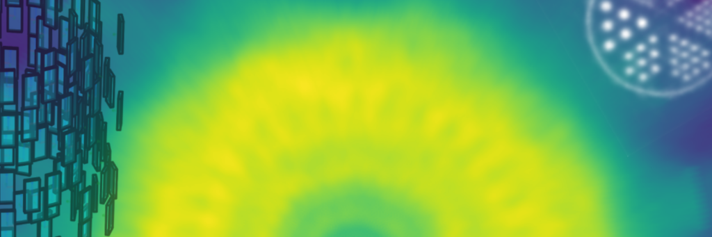
Figure 1: Lightspreads and positioned coincidences visualized for a PET system build at the institute, featuring semi-monolithic slab arrays.
Novel Reconstruction Techniques
Tomographic image reconstruction can be formulated as the solution of an inverse problem governed by the system’s forward projection model. In practice, the accuracy of this model is limited by residual calibration uncertainties, measurement noise, model inaccuracies, and incomplete or limited-angle data. Robust image reconstruction therefore requires methods that combine accurate physical system modelling with data-driven approaches.
Our research focuses on novel reconstruction methodologies that combine physics-based modelling with modern machine learning techniques. This includes diffusion models and Deep Image Prior approaches for PET image reconstruction, with a particular focus on limited-angle and incomplete data scenarios, where conventional reconstruction methods often fail or produce strong artefacts.
In addition, we develop generic system matrix and forward model generation methods that support arbitrary detector geometries and crystal topologies, enabling reconstruction for unconventional PET system designs and simplifying the development of new scanner concepts. Another research direction is the incorporation of calibration and positioning uncertainties at the level of LORs directly into the reconstruction model, enabling uncertainty-aware image reconstruction and improved quantitative robustness.
Finally, we work on efficient real-time data streaming and reconstruction frameworks designed for deployment on edge devices and distributed computing systems, enabling live reconstruction, rapid prototyping, and scalable processing pipelines for modern PET systems.
Hybrid Imaging
In PET/MR systems, complementary structural and functional information provided by MRI can be directly incorporated into the PET reconstruction process.
We investigate multimodal reconstruction approaches that utilize MR information to guide image formation, incorporate anatomical priors, compensate for motion, and estimate physical effects such as attenuation in a data-driven and physics-consistent manner. Our goal is to develop joint reconstruction and modelling techniques that fully exploit the synergy between PET and MRI to improve image quality, quantitative accuracy, and motion robustness.
Figure 2: Schematic illustration of the tomographic imaging modalities investigated at the institute.
Thesis Topics
We offer various thesis topics on this project. We are looking for highly motivated and creative people with a strong background in physics, mathematics, electrical engineering, computer science or related fields. We offer an interdisciplinary working environment where you can gather hands-on experience to the field of PET imaging and image reconstruction.