
Research
Positronen-Emissions-Tomographie (PET)
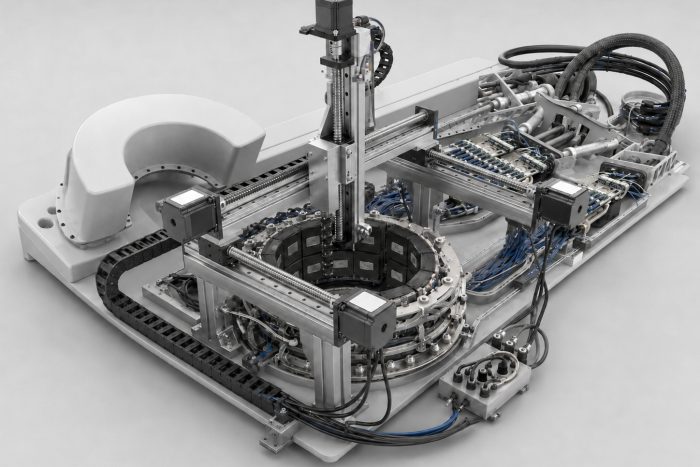
Positron emission tomography shows metabolic processes. A radioactively labelled tracer accumulates in the target tissue; as it decays, two gamma quanta are emitted in opposite directions and registered by a detector ring. The image is formed from around 100 million such coincidences. If the difference in arrival time of the two quanta is also measured, the point of origin along the line connecting them can be localised considerably more precisely.
It becomes particularly interesting when PET and MRI measure simultaneously: functional and anatomical information without registration errors, high soft-tissue contrast, no additional radiation exposure. This requires detectors that neither disturb the magnetic field nor are disturbed by it. That is exactly what we work on, from detector physics through electronics and firmware to reconstruction.
Research Topics

PET Image Reconstruction
Motivation Positron emission tomography (PET) is a functional medical imaging technique that has proven itself in areas such as oncology, cardiology and neurology…

PET/MRI System Development
Motivation Positron emission tomography (PET) is a highly sensitive medical imaging modality with broad application, for example in oncology for tumour diagnosis. PET is based…